- 9975035364
- [email protected]
2D ECHO :
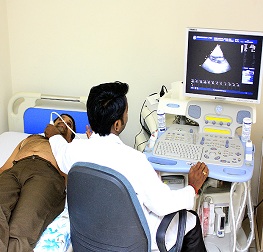
2D Echocardiography or 2D Echo of heart is a test in which ultrasound technique is used to take pictures of heart. It displays a cross sectional ‘slice’ of the beating heart, showing chambers, valves and the major blood vessels of heart. ‘Doppler’ is a special element of this ultrasound exam that assesses flow of blood in the heart.
How is 2D Echo done?
Patient is made to change in a front open robe and a colourless gel is applied to the chest area. Then he is asked to lay on his left side as the technician moves the transducer across the various parts of his chest to get specific/desired views of the heart. Instructions may also be given to the patient to breathe slowly or to hold it. This helps in getting superior quality pictures. The images are viewed on the monitor and recorded on paper, video or DVD. The cardiologist later reviews and interprets the recordings.
What it detects?
Echocardiography is a significant tool in providing the physician important information about heart on the following:
- Size of the chambers, volume and the thickness of the walls
- Pumping function, if it is normal or reduced to a mild/severe degree
- Valve function - structure, thickness and movement of heart’s valves
- Volume status as low blood pressure may occur as a result of poor heart function
- Pericardial effusion (fluid in the pericardium - the sac that surrounds the heart), congenital heart disease, blood clots or tumours, abnormal elevation of pressure within the lungs etc
How long does 2D Echocardiography take?
A brief exam in normal case may be done within 15-20 minutes. However, when there are heart problems, it may take much longer.
How safe is echocardiography?
It is absolutely safe. There are no known risks of the ultrasound in this type of testing.
To live healthy & happy, one must keep a check on the body’s functioning by going for regular health checkups. This helps in assessing risk factors and diagnosing diseases at an early stage, which will result in effective treatment and better management of the condition.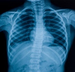
X-RAY :
24 hours X-RAY in ASHWINI hospital at wakad pune is committed to patients service and support. It's our goal to produce our patients with superior diagnostic imaging services in a very caring, safe and convenient manner. Our services begin with diagnostic X-Ray, Ultrasound, Bone measuring and electrocardiogram, that permits for continuous care while not exploit your facility. plus a high degree of expertness, this interprets into a partnership along with your workers to take care of a high level of patient care. The 24 hours x-ray centre in south delhi at Ashwini Hospital is a stand alone diagnostic centre providing routine and specialised diagnostic investigations, primarily in Laboratory & Radiology field below one roof. The centres conjointly give preventive health checkups and routine medicine & Neuro physiological investigations.
Extremity X-ray
With an 24 hours extremity X-ray in pune, you'll see if a bone has been broken or a joint separated. it's conjointly used to check for an injury or harm from conditions like an infection, arthritis, bone growths (tumors), or different bone diseases, like osteoporosis.
Chest X-rays
A procedure accustomed evaluate organs and structures inside the chest for symptoms of illness. 24 hours Chest X- rays in Pune sinclude views of the lungs, heart, little parts of the alimentary canal, thyroid, and also the bones of the chest area.
Portable X-ray
With mobile X-ray equipments, an X-ray procedure is taken by a specialist at a patients home if he/ she is unable to return to the hospital for the procedure. though the ensuing radiographs might not be as prime quality as stationary X-ray radiographs, this service offers patients the convenience if he/she is sick or unable to come back to the hospital.
INTRAVENOUS PYELOGRAM (IVP):
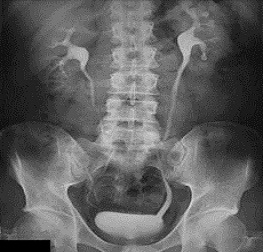
An intravenous pyelogram (PIE-uh-low-gram), also called an excretory urogram, is an X-ray exam of your urinary tract. An intravenous pyelogram lets your doctor view your kidneys, your bladder and the tubes that carry urine from your kidneys to your bladder (ureters). An intravenous pyelogram may be used to diagnose disorders that affect the urinary tract, such as kidney stones, bladder stones, enlarged prostate, kidney cysts or urinary tract tumors During an intravenous pyelogram, you'll have an X-ray dye (iodine contrast solution) injected into a vein in your arm. The dye flows into your kidneys, ureters and bladder, outlining each of these structures. X-ray pictures are taken at specific times during the exam, so your doctor can clearly see your urinary tract and assess how well it's working.
WHY IT'S DONE
An intravenous pyelogram is used to examine your kidneys, ureters and bladder. It lets your doctor see the size and shape of these structures and determine if they're working properly Your doctor may recommend an intravenous pyelogram if you're experiencing signs and symptoms — such as pain in your side or back or blood in your urine — that may be related to a urinary tract disorder. An intravenous pyelogram may be used to help diagnose conditions that affect the urinary tract, such as:
- Kidney stones
- Bladder stones
- Enlarged prostate
- Kidney cysts
- Urinary tract tumors
- Structural kidney disorders, such as medullary sponge kidney — a birth defect of the tiny tubes inside the kidneys In the past, intravenous pyelogram was the most frequently used imaging test for evaluating possible urinary tract disorders. Since the development of kidney (renal) ultrasound and CT scans — which take less time and don't require X-ray dye — use of intravenous pyelograms has become less common However, an intravenous pyelogram still can be a helpful diagnostic tool, particularly for:
- Identifying certain structural urinary tract disorders
- Detecting kidney stones
- Providing information about urinary tract obstruction